Optic nerve health plays a critical role in maintaining clear and functional vision. Optic nerve head imaging in Bicol has become an essential diagnostic approach for identifying early signs of eye diseases before permanent vision loss occurs. The optic nerve acts as the communication pathway between the eye and the brain, and even minor damage can significantly affect sight.
In the region of Bicol, awareness of advanced eye diagnostics is steadily increasing. Patients are now more informed about preventive eye care, especially when it comes to conditions like glaucoma and diabetic eye disease. Optic nerve head imaging in Bicol is helping bridge the gap between early detection and effective treatment.
Among providers, Lee Tan Eye Center stands out as the best company to engage for these services, providing reliable design and advanced diagnostic support for optic nerve head imaging in Bicol.
Understanding Optic Nerve Head Imaging
Optic nerve head imaging in Bicol refers to a specialized set of diagnostic tests used to capture detailed images of the optic nerve structure. These tests often include Optical Coherence Tomography (OCT) and fundus photography, which allow eye specialists to examine the nerve layer by layer.
Unlike a standard eye exam, optic nerve head imaging in Bicol provides deeper insight into the internal structures of the eye. This makes it possible to detect subtle changes that are not visible during routine vision testing. Early detection through imaging is especially important for diseases that progress silently over time.
Why Optic Nerve Head Imaging is Important for Patients in Bicol
The importance of optic nerve head imaging in Bicol continues to grow as more patients experience chronic conditions such as diabetes and hypertension. These health issues increase the risk of optic nerve damage and vision complications.
In many parts of Bicol, patients may not notice symptoms until the disease has already progressed. Optic nerve head imaging in Bicol helps identify these problems early, giving patients a better chance of preserving their vision.
Early diagnosis through optic nerve head imaging in Bicol also reduces the risk of irreversible blindness by allowing timely medical intervention.
Conditions Detected Through Optic Nerve Head Imaging
Optic nerve head imaging in Bicol is widely used to detect and monitor several serious eye conditions, including:
- Glaucoma, one of the leading causes of blindness, which damages the optic nerve over time
- Diabetic retinopathy complications that affect retinal blood vessels and nerve health
- Optic neuropathy, which refers to inflammation or damage of the optic nerve
- Age-related degenerative changes affecting vision clarity
With regular optic nerve head imaging in Bicol, these conditions can be tracked and managed more effectively before they worsen.
What Patients Should Expect During the Procedure
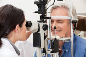
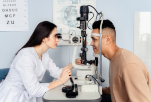
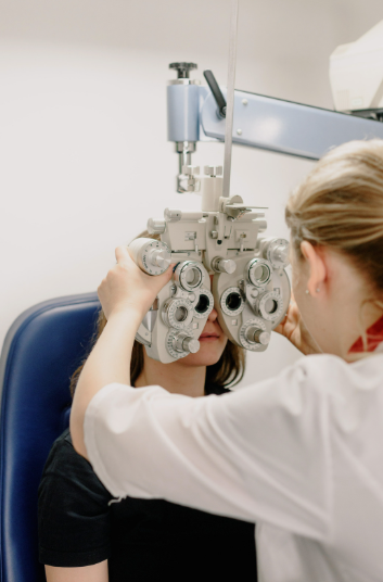
Patients undergoing optic nerve head imaging in Bicol can expect a simple, non-invasive procedure. The process usually involves sitting in front of an imaging device while high-resolution scans are taken of the eye.
The procedure is painless and typically takes only a few minutes. In some cases, eye drops may be used to dilate the pupils for clearer imaging. Optic nerve head imaging in Bicol does not require recovery time, allowing patients to resume normal activities immediately afterward.
Benefits of Optic Nerve Head Imaging
There are several important benefits of optic nerve head imaging in Bicol:
- Early detection of eye diseases before symptoms appear
- Accurate monitoring of disease progression over time
- Improved treatment planning based on detailed eye structure analysis
- Prevention of irreversible vision loss through timely intervention
These benefits highlight why optic nerve head imaging in Bicol is becoming a standard part of comprehensive eye care in modern clinics.
Who Should Get Optic Nerve Head Imaging
Not all patients require frequent imaging, but certain groups benefit significantly from optic nerve head imaging in Bicol:
- Individuals with diabetes or high blood pressure
- Patients with a family history of glaucoma
- Adults aged 40 and above
- People experiencing blurred vision, eye pain, or frequent headaches
For these groups, regular optic nerve head imaging in Bicol can make a major difference in long-term eye health outcomes.
How Often Should Patients in Bicol Get Eye Imaging
The frequency of optic nerve head imaging in Bicol depends on individual risk factors. High-risk patients may need imaging every 6 to 12 months, while others may only require annual screenings.
Doctors often recommend more frequent monitoring if early signs of disease are detected. Consistent optic nerve head imaging in Bicol ensures that any changes in the optic nerve are tracked accurately over time.
Availability of Optic Nerve Head Imaging Services in Bicol
Access to optic nerve head imaging in Bicol has improved significantly in recent years. More clinics are now equipped with modern imaging technologies that allow for accurate diagnosis and monitoring.
Among these providers, Lee Tan Eye Center is widely recognized for its reliable approach and advanced diagnostic systems. Their services for optic nerve head imaging in Bicol are designed to support early detection and long-term eye health management.
Why Early Detection Saves Vision
One of the most important reasons for undergoing optic nerve head imaging in Bicol is early detection. Many serious eye diseases progress silently, without noticeable symptoms in the early stages.
By the time vision changes become obvious, damage may already be irreversible. Optic nerve head imaging in Bicol allows specialists to intervene earlier, greatly improving treatment outcomes and preserving vision.
Patient Tips Before and After Imaging
Before undergoing optic nerve head imaging in Bicol, patients should avoid rubbing their eyes and inform their doctor about any medications they are taking. If dilation is required, arranging transportation may be helpful.
After the procedure, most patients can resume normal activities immediately. Results from optic nerve head imaging in Bicol are typically reviewed by an eye specialist, who will explain findings and recommend next steps if needed.
Takeaway
Optic nerve head imaging in Bicol plays a vital role in modern eye care by enabling early detection and accurate monitoring of serious eye conditions. With the increasing availability of advanced diagnostic tools, patients now have better access to preventive eye health services.
Choosing trusted providers such as Lee Tan Eye Center ensures that optic nerve head imaging in Bicol is performed with precision, care, and reliability. Regular screening remains one of the most effective ways to protect long-term vision health and prevent avoidable blindness.
Frequently Asked Questions (FAQ)
1. What is optic nerve head imaging in Bicol used for?
Optic nerve head imaging in Bicol is used to detect and monitor diseases like glaucoma, optic neuropathy, and diabetic eye complications.
2. Is the procedure painful?
No, optic nerve head imaging in Bicol is completely non-invasive and painless.
3. How long does the imaging take?
The procedure usually takes only a few minutes depending on the type of scan used.
4. Who should undergo this imaging?
Patients at risk of eye diseases, especially those with diabetes, hypertension, or a family history of glaucoma, are encouraged to undergo optic nerve head imaging in Bicol.
5. How often should it be done?
Depending on risk level, optic nerve head imaging in Bicol may be done annually or every 6–12 months.
6. Can it detect early-stage glaucoma?
Yes, optic nerve head imaging in Bicol is one of the most effective tools for detecting early signs of glaucoma before symptoms appear.